Pictured above: 3D rendering of a single congenital left gastric portosystemic shunt (ventral view), 3D rendering of a right ectopic ureter (lateral view)
CT is a tomographic imaging technique that uses x-ray beam to create high-resolution, thin-section anatomical images. During CT, the animal is placed on a table which moves through the circular opening of the CT scanner (the gantry). X-rays are generated by a high-power x-ray tube in the gantry; after the x-rays travel through the patient, their attenuation is measured by detectors located on the opposite side of the gantry. Attenuation values are translated into CT numbers (or Hounsfield units, HU) and converted to shades of gray that are displayed as an image. The sensitivity of CT to subtle difference in x-ray attenuation is 10 times higher than conventional radiography: this means that CT identifies bone lysis and production earlier that radiography. In addition, CT allows evaluation of osseous, pulmonary and soft tissue structures with a single scan, by manipulating the image display. Further, cross sectional images greatly facilitate evaluation of area of complex anatomy, such as joint, the thorax and the skull.
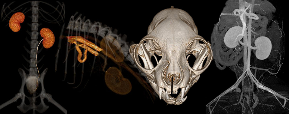
There are many indications to use CT in small animals; common applications include evaluation of nasal disease, ear disease, pulmonary pathology, vascular abnormalities, and musculoskeletal disorders. Our current CT scanner, a Philips Brilliance 40-channel multidetector CT interfaced with an image workstation, offers ultra-fast scan times, and generates images with sub-millimeter resolution. This translates in shorted anesthesia time (and even allows some scans to be performed using sedation only), while enhancing our board-certified radiologists’ diagnostic accuracy.
