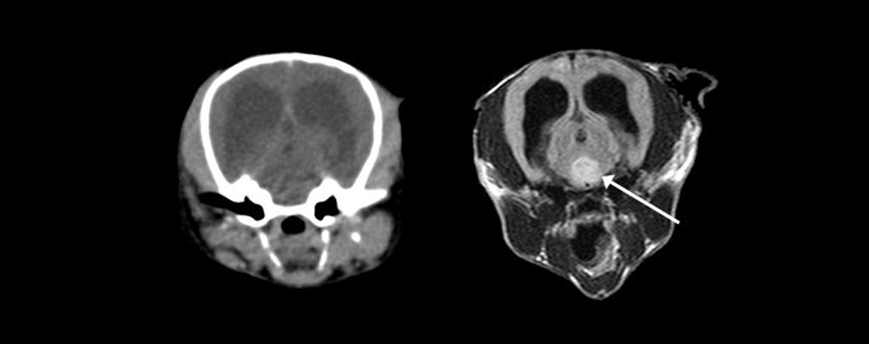
Pictured above: These are computed tomographic and magnetic resonance images of the brain in the same dog. The MR image (right) shows a lesion in the brain parenchyma (arrow), which is not evident on the CT image (left)
MRI is an imaging method based principally upon sensitivity to the presence and properties of water (more precise: mobile hydrogen protons). “Water” is present in abundance in the body, making MRI a useful tool in assessing physiologic and pathologic processes.
In MR terminology “water” describes both “free water” (liquids like urine, CSF, cystic fluids) and “bound water” (intra- and extracellular fluid in tissues). Pathologic processes change the water content and properties in tissues. Examples include cysts or abscesses (i.e. cavities containing free water), edema (i.e. increase in water content of tissue), inflammation and neoplasia (i.e. presence of cells that normally would not be present).
MRI is very sensitive in picking up these abnormalities in water distribution and composition.
Images are created by subjecting the patients to a magnetic field and monitoring the behavior of tissues in this environment. MRI does not involve radiation, and is therefore considered a very safe imaging modality.
